Raman spectroscopy for the identification of isocitrate dehydrogenase (IDH) mutated glioblastomas.
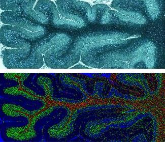 Image credit: Own Work
Objectives:
Determining IDH status of glioblastomas is critical for histological diagnosis and clinical decision-making. Raman spectroscopy probes the unique molecular vibrations of a sample to accurately characterise its molecular composition. We investigate the feasibility of using Raman spectroscopy to differentiate between IDH1 positive and negative tumours.
Design:
Spectroscopic analysis of neuropathological samples.
Subjects:
40 glioblastoma formalin fixed samples classified into either IDH1þ or IDH1- using standard immunohistochemistry IDH1(R132H) stain. 3 additional samples with rare IDH1 mutations (R132G) were also used. Methods: Spectral maps of 1mm2 were acquired from two areas on each sample using a Raman microscope configured with a 785nm excitation laser source. Acquisition time less than 60 minutes per map.
Results:
72 spectral maps (145,800 spectra) from 36 samples (18 IDHþ and 18 IDH-) were included in the analysis. 4 hypocellular samples were excluded. Principal component analysis (PCA) demonstrated good separation of the IDH1þ and IDH1- groups. A PCAlinear discriminant analysis classification model demonstrated 97% sensitivity and 90% specificity for predicting the presence of an IDH1 mutation. We also demonstrated similar accuracy when classifying rare IDH1 mutations into the IDH þ group.
Conclusions:
Raman spectroscopy can accurately and rapidly distinguish IDH1 mutated glioblastomas from their IDH1 negative counterparts. Further work is currently being undertaken on fresh, intraoperative, tumour samples.