Raman spectroscopy for the identification of isocitrate dehydrogenase (IDH) mutated glioblastomas.
Investigated the feasibility of using Raman spectroscopy to differentiate between IDH1 positive (IDH1 + ) and negative (IDH1 - ) tumours through classification modelling.
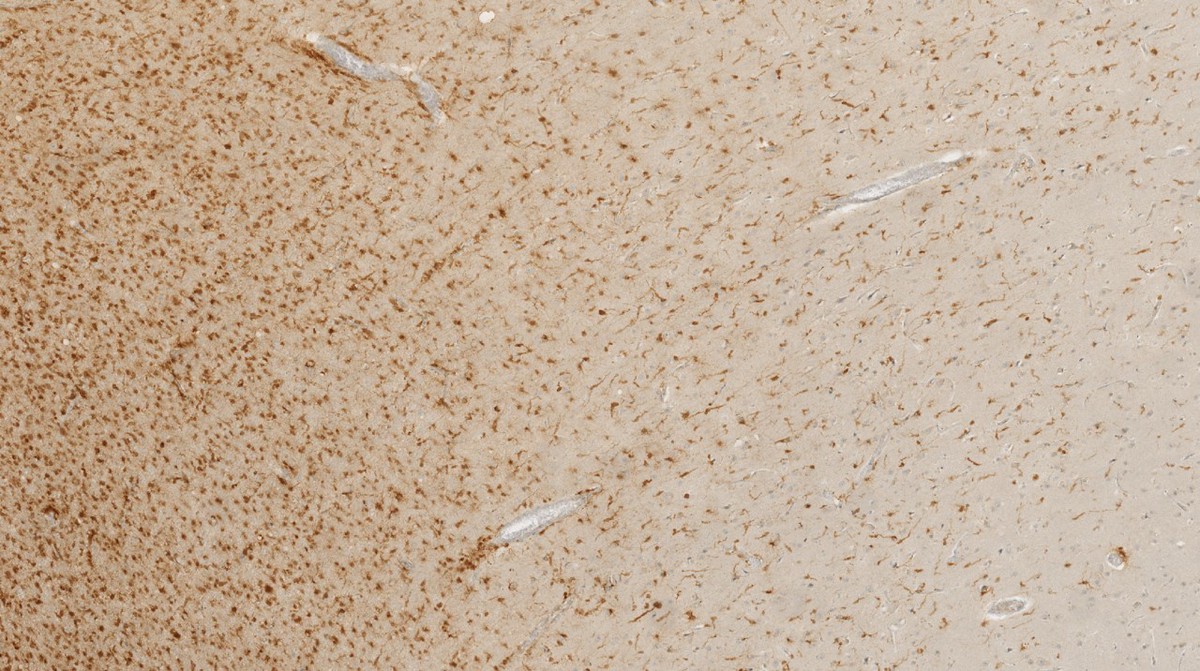 Image credit: Own Work
Introduction: Raman spectroscopy is an emerging biophotonic tool for the identification of disease. By probing the unique molecular vibrations that depend on the composition and structure of samples, it provides a wealth of information on a cellular and molecular level of both solid and liquid specimens without the use of external agents such as dyes, stains or radioactive labels. Glioblastomas can be stratified by the presence or absence of mutations in isocitrate dehydrogenase (IDH) 1 or 2. Determination of IDH status is critical for histological diagnosis and clinical decision-making with evidence that the presence of IDH mutation confers better prognosis and a better response to chemotherapy. Mutation-positive tumours accumulate high concentrations of D-2-hydroxyglutarate, resulting in metabolic and epigenetic changes which we hypothesize will be reflected in a change in Raman scattering. In this study, we investigate the feasibility of using Raman spectroscopy to differentiate between IDH1 positive (IDH1 + ) and negative (IDH1 - ) tumours through classification modelling.
Method: Forty biopsies of glioblastoma (WHO grade IV) in patients under the age of 40 years were classified into either IDH1+ (n=20) or IDH1- (n=20) using IDH-1(R132H) and BCAT-1 immunohistochemistry. 6µm thick sections were prepared from formalin fixed paraffin embedded specimens. Based on the hematoxylin and eosin stained contiguous section, high tumour density areas in the unstained sections were identified and subjected to analysis. Raman maps of ca. 1mm 2 were acquired from up to two locations on each sample using an inVia Raman microscope (Renishaw, UK) configured with a 785nm excitation laser source.
Results: 30 spectral maps (60,325 spectra) from a total of 16 biopsy samples (8 IDH+ and 8 IDH-) have been collected to date. Principal component analysis (PCA) demonstrated good separation of the IDH1+ and IDH1- groups. A PCA-linear discriminant analysis classification model demonstrated 96.98% sensitivity and 90.1% specificity for predicting the presence of an IDH1 mutation.
Conclusions: The results demonstrate the feasibility of using Raman spectroscopy to accurately distinguish IDH1 mutated glioblastomas from their IDH1 - counterparts. Raman spectroscopy could provide a powerful tool to aid neuropathological diagnosis and genetic classification of glial tumours in the future.
Acknowledgements: This work was supported by Cancer Research UK (CRUK) grant number C38302/A17319, and the NIHR Oxford Biomedical Research Centre
Published poster in Neuro-Oncology (Volume 19, Issue Supplement 1, Janurary 2017, Pages 14-15) in addition to the original abstract published in October 2016.