The BigMac dataset - linking tissue microstructure with diffusion MRI signals throughout the brain
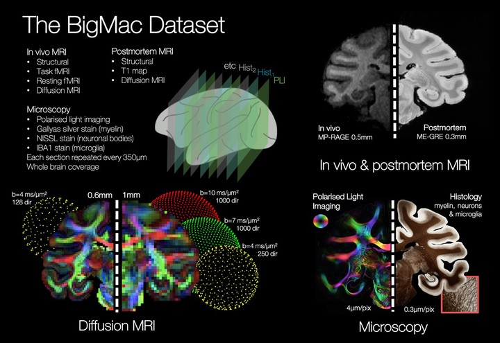 Image credit: From Abstract
Our ability to characterise the human connectome has been greatly advanced by the scientific community’s access to big data. Big data can be population based (to examine both inter- and intra-subject variability in e.g. Biobank1 or the HCP2) or can aim to characterise a single connectome in exquisite detail3. Here we present the BigMac dataset which combines in vivo MRI with postmortem ultra-high angular resolution diffusion imaging (ultra-HARDI) and multi-contrast microscopy data in a single macaque brain. Once published, the dataset will provide a platform from which we can interconnect microstructural features of the tissue with MRI signals throughout the brain. Figure 1 shows some example images from the BigMac dataset. Importantly, the BigMac dataset includes in vivo MRI data. This provides an excellent opportunity to link high-quality postmortem MRI and microscopy data (detailed structural connectivity) to typical in vivo data (structural and functional connectivity) within the same brain. Post sacrifice, an comprehensive MRI dataset (~270hrs scanning time) was acquired which includes both high resolution (0.3mm isotropic) structural images and extensive diffusion MRI data at two spatial resolutions (0.6mm and 1mm isotropic), b-values of up to 10ms/µm2 and 1000 gradient directions in the outer two shells (‘ultra-HARDI’). The ultra-HARDI data allows us to both characterise the diffusion signal in great detail and retain dense sampling on an arbitrary 2D plane for direct comparison with 2D microscopy data4. Furthermore, with 1000 gradient directions we see more biologically plausible reconstructions of technically challenging white matter tracts such as longitudinal fasciculus II4. After scanning, the brain was sectioned along the entire rostral-caudal axis with consecutive slices processed for polarised light imaging5 (to visualise the orientation of myelinated fibres) or histological staining of myelin, neuronal bodies or microglia. These sections are then imaged at high resolution (0.3-4 µm/pixel) and co-registered to the MRI6. The BigMac dataset is interesting from both an anatomical and methodological viewpoint. The densely sampled multi-contrast microscopy can be used to examine both the myelo- and cyto-architecture in great detail and develop novel white matter / grey matter atlases or parcellations. For those interested in diffusion modelling, the comprehensively sampled diffusion MRI space can be used to drive protocol optimisation, advanced biophysical modelling of the tissue microstructure and direct validation of many current and future diffusion MRI models. Finally, the BigMac dataset will support many machine learning algorithms which jointly model MRI and microscopy data to, for example, map quantitative imaging biomarkers to specific features of the tissue microstructure.